
Advanced Imaging
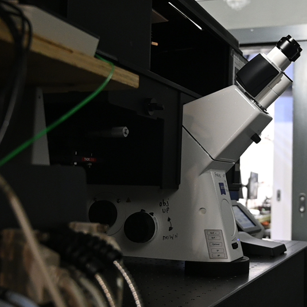
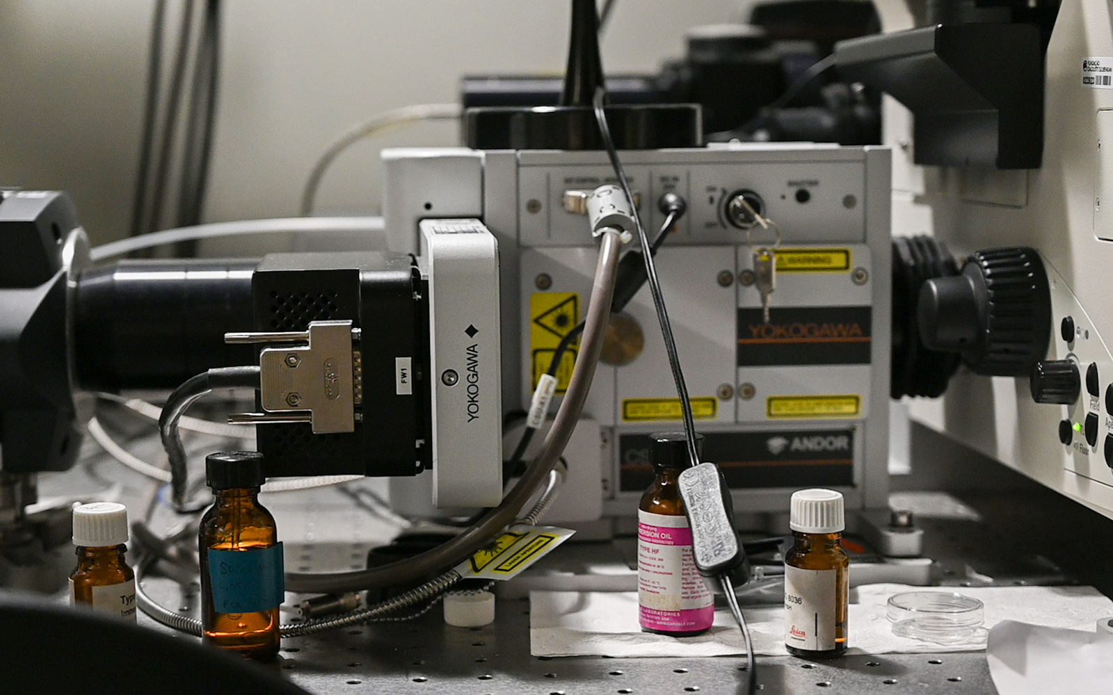
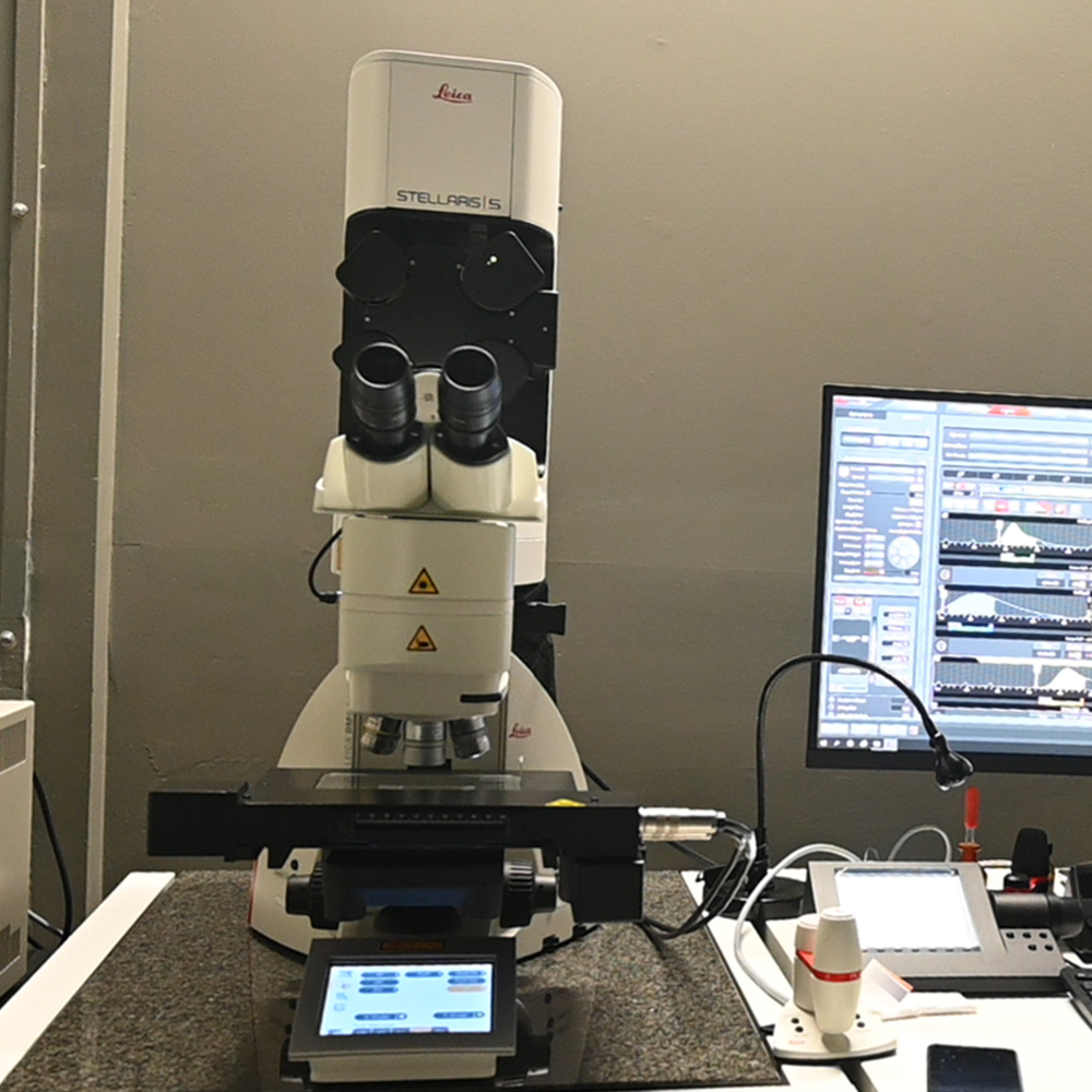
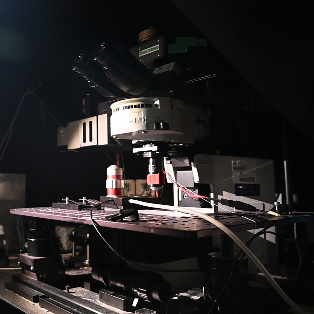
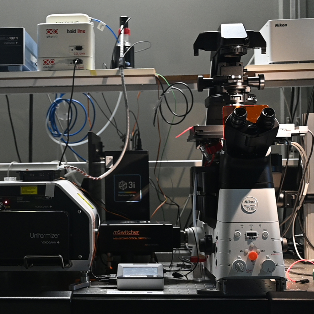
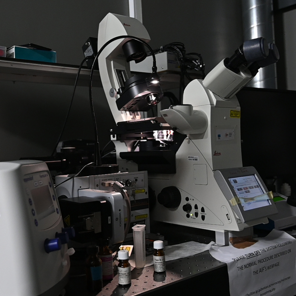
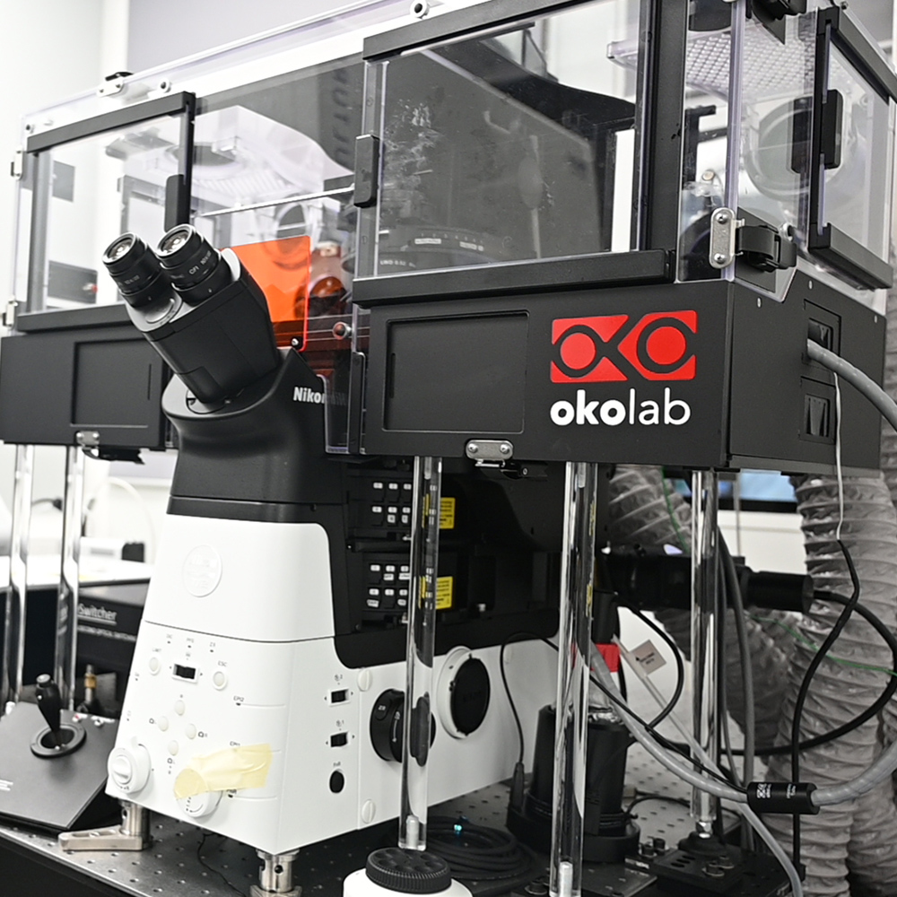
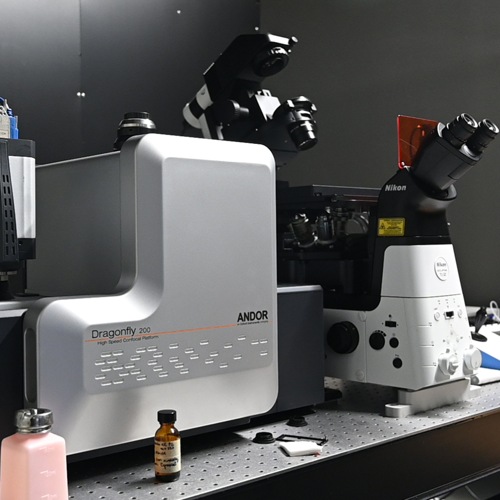
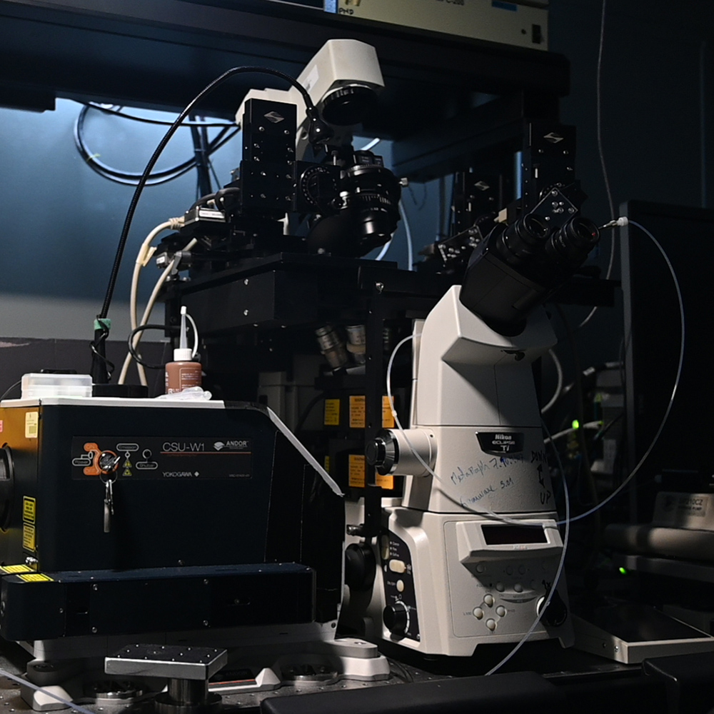
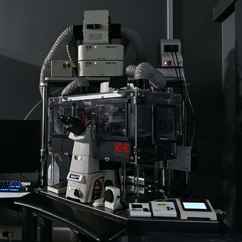
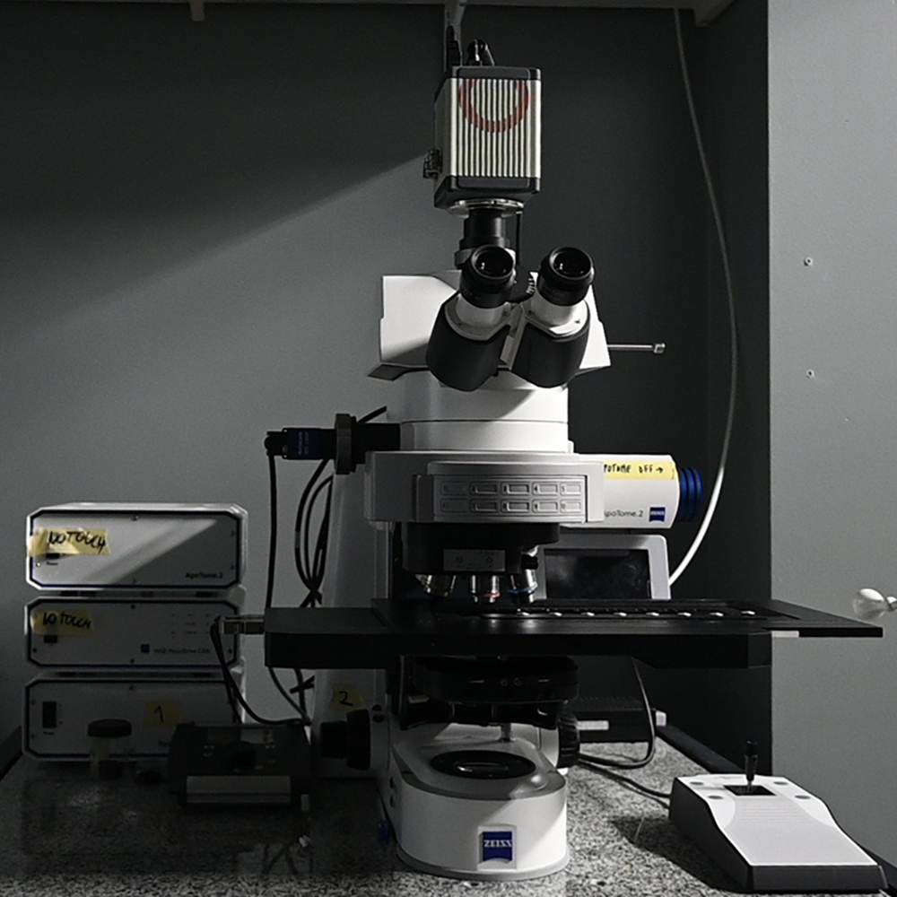
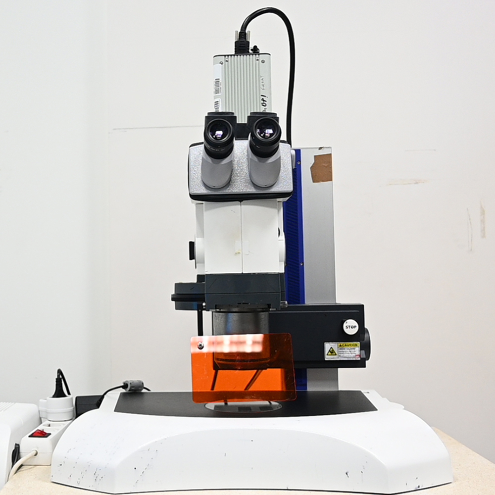
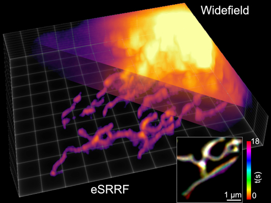
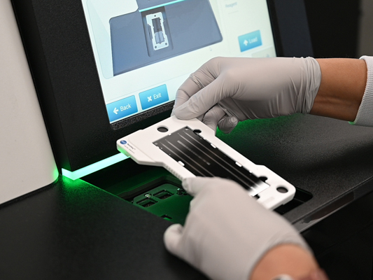
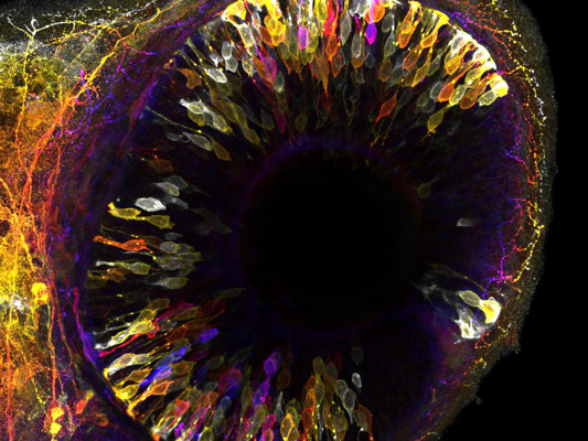
The IGC’s Advanced Imaging Unit offers state-of-the-art light microscopy and bioimaging services, housing 16 high-end systems with confocal/2photon, high-throughput, super-res & mesoscopy (light-sheet & OPT). Services include sample prep., and training with instrumentation & bioimage analysis (high-end workstations available with commercial & free software). The AIU is open-access and serves the IGC, academia and companies.
Location: Bartolomeu Dias wing
Contact: +351 214 464 539 @: [email protected]
Users of the AIU should observe the “General IGC Facilities usage” terms which can be consulted here (requires login in Agendo).
The IGC’s “code of conduct” also contains relevant information about the expected conduct at the facility.
How to avoid negligent use of the AIU?
Any negligent use should be promptly reported (by users and staff) to the facility head or staff, which will evaluate whether the user should be denied future booking and use. Agendo allows reporting “incidents” that should be used when such problems are detected. Examples of what may be considered negligent use:
- Forgetting to turn off the instruments or critical components such as fluorescent lamps & lasers, when no one else is booked after (check the calendar or ask staff before leaving),
- Failure to clean or report dirty optics or system malfunction to staff
- Repeatedly arriving late, skipping or deleting booked sessions without providing an explanation; using equipment without, or past, the time booked without informing the staff.
- Repeatedly booking on behalf of others.
- Attempting to use the system without training and previous authorisation from staff
- Attempting to alter the characteristics or functionality of the instruments, or parts, without previous training AND consent from the staff (eg removing/replacing objectives)
- Installing any software or connecting external disks/USB sticks to workstations
- Making use of the workstations and internet access for purposes other than checking protocols, system booking, acquiring, processing/analysing & transferring image datasets.
- Falsification of bioimage results or purposeful misrepresentation of bioimaging data.
What may constitute an intellectual contribution from AIU staff?
Granting equipment access, training and providing consumables, per se, DO NOT warrant co-authorship in publications from any of the facility staff.
Support from staff with acquisition of raw datasets (image data acquired “as is”, without pre-processing), opening and converting to other file generic formats, basic process & analysis procedures (eg, contrast enhancement, 2D measurements, basic segmentation and particle analysis) also DO NOT constitute a significant contribution that would warrant co-authorship in publications. Help with deconvolution and other basic forms of data restoration or pre-processing (eg. cleaning noise, linearization or background removal)vare also not considered significant intellectual contributions.
The following may constitute an intellectual contribution, and should be discussed (either in advance or a posteriori) with the facility head and staff member providing support:
- Implementation of novel (or uncommon/untested) protocols for sample preparation, image acquisition, processing and/or analysis.
- Custom-building of hardware or software (“Technical Development”)
- Obtaining a substantial amount of the images used in a publication (or the ones presented).
- Complex image feature enhancement or extraction, 3D/4D image segmentation/tracking, complex measurements (eg, those not available in ImageJ’s “particle analysis” or “set measurements”)
- Preparation of manuscript plates/figures or text (even if later they are altered by the researchers) - note that suggestions for Material & Methods are available at the website.
- Preparation of complex macros/scripts for automation of processing & analysis
- Performing statistical tests on bioimage derived data
- Preparation of complex 3D renderings & animations/illustrations
- Processing, segmenting and analysis of large volumes of data
- Implementation of tools not common in a facility or for which the staff are not trained
In these cases, the facility staff should be consulted before publication to make sure the methods/results are properly described and acknowledged. When co-authorship is warranted, the senior author should obtain the agreement of the imaging staff member regarding his/her contribution and the contents of the publication and be allowed to review & edit the relevant work being submitted to publication. When deemed necessary, the Facility users’ committee will be consulted to provide assistance in evaluating the significance of intellectual contributions.
Payment of imaging services is always due, even when co-authorship is warranted.
Acknowledging the AIU in publications?
All AIU users are requested to acknowledge the support of the AIU in their publications and communications involving any of our services, in order to guarantee future funding of the facility. There is a specific acknowledgement for each instrument, so please consult the appropriate SOP/user guide. In all cases, it should at least include the following: “We thank the support of IGC's Advanced Imaging Facility (AIU), especially [insert name of staff member(s) who helped], funded by PPBI-POCI-01-0145-FEDER-022122 (Lisboa 2020/FEDER/FCT; Portugal).
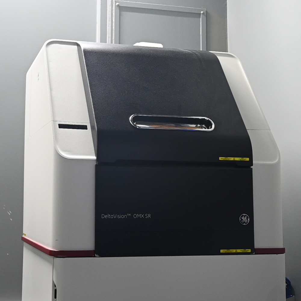
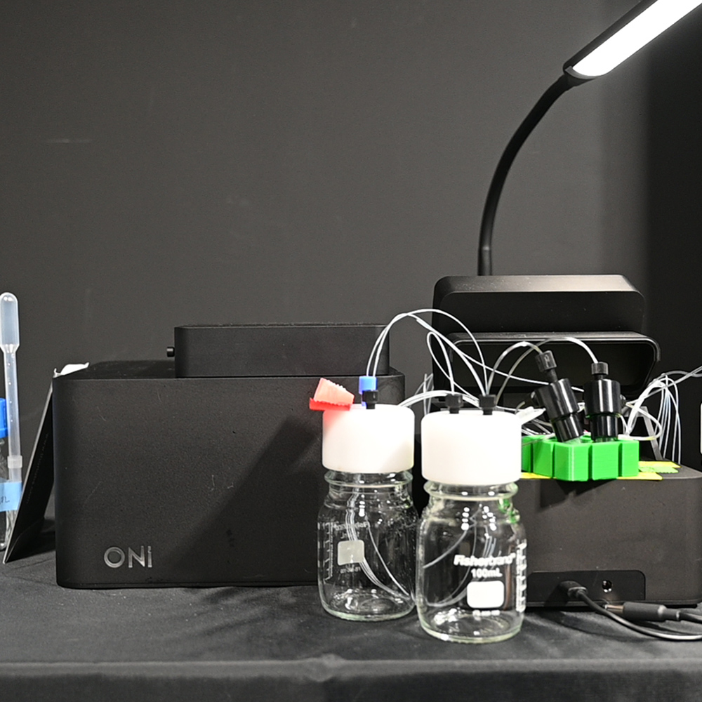
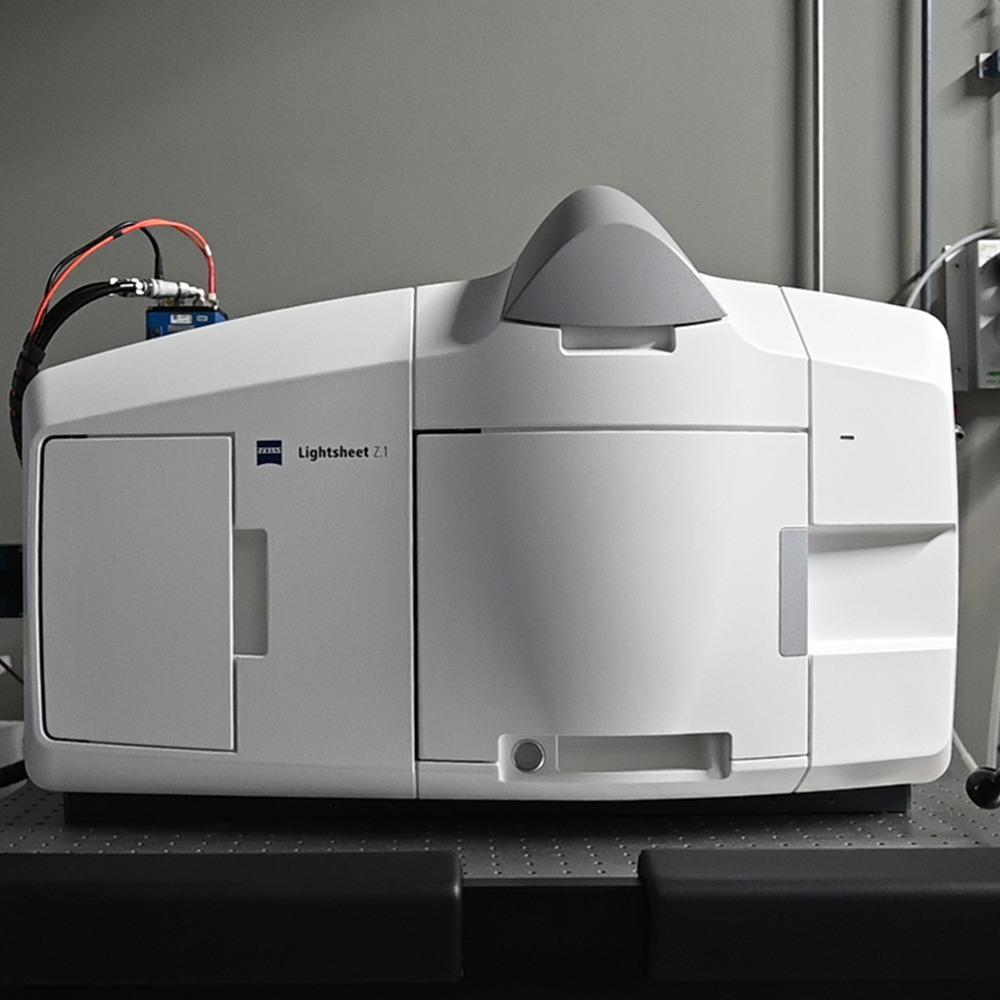
EQUIPMENT
SERVICES & TECHNIQUES
Experimental design for imaging, cell / tissue staining; sample preparation and tissue clearing.
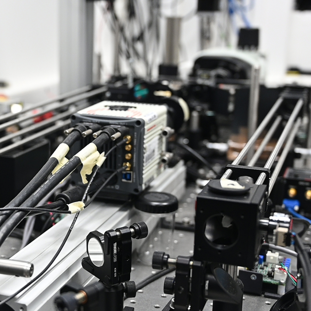
Diagnostics, repair and building optical systems.
Pending availability of staff and project demand.
ImageJ, QuPath; Imaris, Huygens, Amira and others - for more information please contact.
Laser scanning & spinning disk; Airyscan, Lightning & SORA available.
Intravital, SHG possible.
Multiposition; tile & stitching; live-imaging; well-plates.
Up to 8 per run.
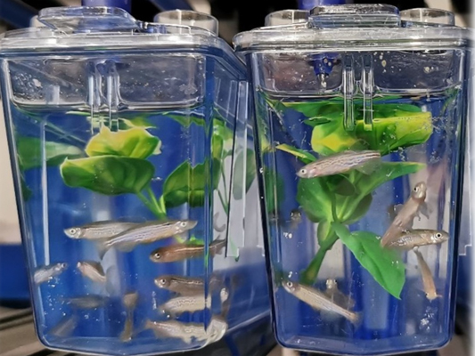
Filming of embryos or organoids in vivo or acquisition of volumetric 3D images of meso and macro cleared samples, by light-sheet (fluorescent) or OPT (fluorescent or unlabeled).
SIM and Apotome.
STORM/PALM/TIRF.
Micro & meso - for CLEM see EMF.
Ablation, FRAP, FLIP, FRET(AB/SE).
PROJECTS
Referência do projeto: PTDC/BII-BTI/32375/2017
Código do projeto: 02/SAICT/2017
Objetivo principal: OT 1 – Reforçar a investigação, o desenvolvimento tecnológico e a inovação
Localização do projeto (NUTS II): Lisboa (100%)
Entidade Beneficiária:
Fundação Calouste Gulbenkian - Instituto Gulbenkian de Ciência
Data de aprovação: 23-03-2018
Data de início: 01-10-2018
Data de conclusão: 30-09-2021
Custo total elegível: 238.854,56 EUR
Apoio financeiro publico nacional (orçamento de estado): 238.854,56 EUR
Descrição do projeto:
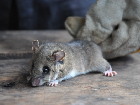
A microTomografia Óptica (OPT) é uma técnica pouco explorada em pesquisa biomédica. A OPT fornece imagens 3D isométricas de amostras grandes que não podem ser visualizadas por microscopia confocal, 2-fotões ou até folha-de-luz. O OPT foi fundamental para a criação das modernas bases de dados de desenvolvimento de vertebrados e, por exemplo, nos esforços do "International Mouse Phenotyping Consortium" para caracterizar fenotipicamente milhares de linhas de ratos knockout, sendo a ferramenta ideal, quer em termos de qualidade de imagem quer de rendimento (Ruparelia etal2014; Dickinson etal2016). Introduzimos o primeiro scanner OPT totalmente aberto, integrado como parte da plataforma OpenSpin que opera sistemas light-sheet (LSM) e OPT (Gualda et al 2013). OPenT é um spinoff do OpenSpin, já com +4 publicações (Ruparelia et al2014; Gualda et al2014; Zeng et al2015; Felix et al2016), 2 bases anatómicas on-line, 1 prémio internacional e 13 laboratórios internacionais que pediram suporte para construir aas suas próprias versões. Propomos a integração de LSM+OPT num mesoscópio multimodal de alta capacidade, a funcionar na Unidade de Imagem Avançada do IGC, que exploraremos para caraterizar embriões mutantes de murganhos e larvas de Danio, dois importantes modelos experimentais de doença humana. Esta técnica irá acelerar a descoberta, proporcionando acesso simplificado e totalmente aberto para experiências complexas de imagem 3D a órgãos ou organismos inteiros, a laboratórios que de outra forma não dispõem do financiamento ou infraestrutura necessários.
O prazo de execução do projeto OPenT foi prolongado até 30 de setembro de 2022.
FUNDING

USEFUL RESOURCES
Microscopy Primer (basics)
COLife & friends bioimaging basics 2021 webinars
FPbase (fluorescence spectra)
(fluorescence) Spectraviewer
Biii.org (bioimage analysis resources)
Sea4Bia (scripts/macros from PPBI facilities)
NEUBIAS Academy @Home (Image analysis tutorials)
Confocal-list (forum)
Image.Sc (forum)
PEOPLE

No results were found for your criteria.
PUBLICATIONS
Check the papers here.
MEDIA

No results were found for your criteria.
Funding

Partners
![]()
![]()
![]()
![]()

![]()
![]()