Finding the open road: how neurons migrate in the developing retina

Researchers from the Instituto Gulbenkian de Ciência (IGC), the Max Planck Institute of Molecular Cell Biology & Genetics and the Technische Universität in Dresden, Germany, found a type of cell migration in the retina not known before to be present in the developing nervous system. The study, published in the scientific journal eLife, helps to further understand the different ways neurons use to migrate to ensure the formation of the intricate structure that is our brain, with a special focus on the retina.
The retina is a part of the central nervous system and is responsible for one of our most important senses, vision. Without it, we would not be able to capture images of our environment, which would impair our movements, tasks, and interactions with others. Despite the undeniable importance of this organ, it is not yet fully understood how it is built and maintained.
Neurons in the retina, as well as in other parts of the brain, are usually born at positions different from where they ultimately perform their function. Thus, during development they must migrate to reach their final destinations. How neurons with diverse morphologies migrate successfully in complex and dynamic environments is only beginning to be understood. In most parts of the brain this question is hard to tackle because tissues are not accessible and, consequently, cannot be observed in living organisms. The retina, however, is a region in which this is easier as it is located outside of the embryo. Combining this with the fact that zebrafish embryos are transparent and develop fast generates a perfect system to understand nervous system development where it happens, when it happens.
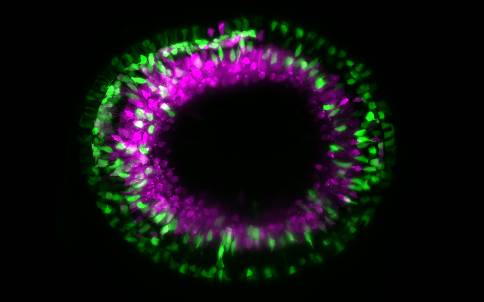
Committed to deepen the understanding of how the retina and, ultimately, the brain develops, an international team of researchers recently studied how a specific type of neuron migrates to form a layer of the zebrafish retina. During this stage of development, the retina is densely packed with other cells, leaving no space for pre-defined paths. So how exactly do these neurons find the open road in the middle of all this traffic?
The researchers found that these neurons tailor their movements to the spatial constraints of the crowded retina by frequently changing their shape and direction. By acquiring a crawling-like type of movement, they can follow flexible trajectories and overcome obstacles to reach their final positions. They also observed that several protrusions extend from their cell body during migration. But whether these protrusions directly drive migration, explore environmental cues or both, remains to be further investigated.
“This type of migration allows the cells to follow the path of least resistance when moving in the densely-packed retina”, explains Caren Norden, principal investigator at the IGC and co-author of the study. “How exactly this path is found by the neurons and whether there are additional factors that help them move forward will be exciting avenues for future studies”, she adds. But this study already left us with some clues regarding how the surrounding environment could influence the neuron’s ability to negotiate space. The researchers found that if they tampered with the deformability of the neighboring cells, many neurons would fail to reach their final positions. This suggests that, to move efficiently, the neurons might need to probe their environment. The authors propose that their protrusions might be involved in this process, given that they simultaneously extended towards multiple directions, even when the neurons were not moving.
Taken together, these findings reveal an additional mode of migration that is integral for the proper development and function of an important region of the central nervous system, the retina. According to the authors, this type of movement will likely also be found in other areas of the brain now that its existence has been revealed. Thus, this and follow up studies will contribute to solve the puzzle of how the retina and the rest of our complex brain is formed correctly to ensure proper function later.
Read Paper
